Differentiating Skin Scanning Analyzers: A Multi-Dimensional Comparison From Principles To Applications
Oct 15, 2025
With the continuous evolution of skin detection technology, skin scanning analyzers have developed into various types and forms, serving different professional needs. Clarifying the differences between them helps practitioners make appropriate choices based on scenarios and functional positioning, thereby improving detection efficiency and service quality.
From the perspective of imaging principles, the main differences lie in the optical technology and resolution depth used. One type is multispectral imaging, utilizing multiple wavelengths of light sources such as visible light, polarized light, and ultraviolet light to acquire comprehensive information from the skin surface to the superficial dermis. It excels at macroscopically quantifying indicators such as pigment distribution, texture roughness, and pore density, and is suitable for daily skin condition assessment and care guidance. Another type is confocal microscopy, based on laser point-by-point scanning and optical sectioning, which can present cellular and even subcellular structural images, such as the arrangement of the stratum corneum and the morphology of collagen fibers. It is more suitable for scientific research or scenarios requiring high-resolution pathological analysis, but has higher requirements for the environment and operation.
From the perspective of detection dimensions, there are two categories: single-modal and multi-modal. Single-modal devices focus on specific indicators, such as capacitive detectors that measure only moisture content or visible light imagers that analyze only pigments. Their advantages lie in their simple structure and low cost, making them suitable for basic screening in primary care institutions. Multimodal fusion devices, on the other hand, integrate two or more detection technologies, simultaneously outputting multi-dimensional data such as texture, pigment, moisture, oil, and sensitivity. They also use algorithms to correlate causes, meeting the refined needs of comprehensive skin management, and are therefore more widely used in mid- to high-end institutions.
From an application perspective, they can be divided into medical clinical and beauty/health applications. Medical clinical devices emphasize diagnostic accuracy and data traceability, often interfacing with medical information systems to assist in skin disease screening and treatment monitoring, with strict requirements for data accuracy and compliance. Beauty/health applications prioritize user experience and intuitive display, with quick testing processes and easy-to-understand graphs, more closely aligned with daily beauty and skincare guidance, emphasizing user-friendly interaction and approachable result presentation.
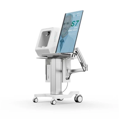
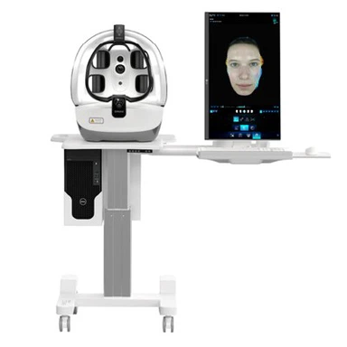
Furthermore, there are also differences in structure and performance. Some devices employ fixed optical paths, offering high stability but limited flexibility; others feature adjustable supports and multi-dimensional positioning systems, better adapting to diverse detection postures and user groups. Performance differences also lie in imaging resolution, data processing speed, and historical data management capabilities, all of which impact the efficiency and continuity of practical use.
In summary, skin scanning analyzers each possess unique characteristics in terms of principle, dimensions, application scenarios, and structural performance. Understanding these differences provides a basis for accurate selection, enabling the equipment to better integrate into different business processes and maximize its effectiveness.