 EN
EN
 AR
AR
 BG
BG
 HR
HR
 CS
CS
 DA
DA
 NL
NL
 FI
FI
 FR
FR
 DE
DE
 EL
EL
 HI
HI
 IT
IT
 JA
JA
 KO
KO
 NO
NO
 PL
PL
 PT
PT
 RO
RO
 RU
RU
 ES
ES
 SV
SV
 TL
TL
 IW
IW
 ID
ID
 SR
SR
 SK
SK
 SL
SL
 UK
UK
 VI
VI
 SQ
SQ
 HU
HU
 TH
TH
 TR
TR
 FA
FA
 AF
AF
 MS
MS
 UR
UR
 BN
BN
 LA
LA

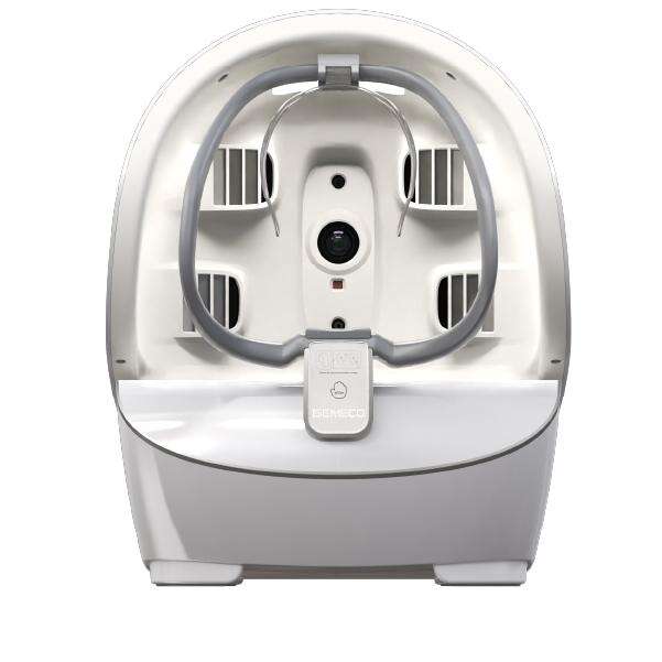
Melasma, a complex pigmentation disorder characterized by irregular, symmetric dark patches on sun-exposed areas, presents a persistent challenge for dermatologists. Its ability to involve both epidermal and dermal layers, combined with its tendency to recur, demands a diagnostic approach that goes beyond visual inspection. MEICET’s Pro-A All-in-One Skin Imaging Analyzer, equipped with multi-spectral imaging, addresses this by dissecting pigment layers, distinguishing subtle patterns, and providing actionable data that transforms vague observations into targeted treatment strategies.
Unpacking Pigment Layers with Multi-Modal Scans
Melasma’s variability—from superficial epidermal involvement to deep dermal penetration—requires a tool that can isolate these layers. The Pro-A’s suite of imaging modes does precisely this:
- Ultraviolet (UV) imaging highlights epidermal melanin, which fluoresces under UV light. In cases of epidermal melasma, this mode reveals bright, well-defined patches that align with visible dark areas, confirming that topical interventions (such as tranexamic acid or kojic acid) may effectively reduce pigment.
- Cross-polarized light (CPL) imaging penetrates beyond the epidermis, visualizing dermal pigment as a distinct gray-blue hue. This is critical for identifying dermal melasma, which often resists topicals and requires more targeted therapies like low-fluence lasers or fractional resurfacing.
- RGB imaging provides high-resolution surface detail, mapping the distribution of pigment across facial landmarks (e.g., cheeks, forehead, upper lip) and confirming symmetry—a hallmark of melasma linked to hormonal or UV triggers.
Consider a patient presenting with symmetric dark patches on the cheeks and forehead. A visual exam alone might suggest “dark spots,” but Pro-A scans reveal nuanced details: UV fluorescence indicating epidermal pigment on the cheeks, CPL showing a diffuse gray-blue pattern on the forehead (dermal involvement), and RGB confirming the patches spare sun-protected areas (e.g., under the chin). This layered data guides a dual treatment plan: topical brighteners to address the epidermal component and gentle laser sessions to target the dermal pigment, avoiding the inefficiency of treating all areas uniformly.
Distinguishing Melasma from Look-Alike Conditions
Misdiagnosis is a significant risk with melasma, as it often mimics post-inflammatory hyperpigmentation (PIH), solar lentigines, or even drug-induced pigmentation. The Pro-A’s pattern analysis helps clinicians draw clear distinctions:
- Melasma typically exhibits bilateral symmetry, worsens with UV exposure or hormonal changes (e.g., pregnancy, oral contraceptives), and involves both epidermal and dermal layers. In CPL mode, its dermal component appears as a “smudged” gray-blue, lacking the sharp borders of PIH.
- PIH arises from prior inflammation (e.g., acne, eczema, or trauma) and fades over time. UV imaging shows it as bright, discrete spots that correspond to the location of previous lesions, with no dermal involvement in CPL mode.
- Solar lentigines (age spots) develop in sun-exposed areas, appear as well-circumscribed dark spots, and show consistent UV fluorescence with no dermal pigment—responding well to targeted laser treatments that melasma might resist.
By correlating these patterns with clinical history, the Pro-A ensures accurate diagnosis. For example, a patient with a history of dark patches that worsened during summer and improved with sunscreen would have Pro-A scans confirming melasma: symmetric distribution, mixed epidermal-dermal involvement, and no link to prior inflammation—ruling out PIH and guiding UV protection and hormonal adjustments alongside topical treatments.
Monitoring Treatment Response Over Time
Melasma’s recurrence risk demands long-term monitoring to adjust therapies and prevent flare-ups. The Pro-A’s follow-up capabilities provide objective metrics to track progress:
- UV intensity measures changes in epidermal pigment. Scans showing diminished fluorescence in previously bright areas confirm that topical brighteners are working, justifying continued use. Conversely, persistent UV brightness signals the need to escalate to more intensive treatments (e.g., chemical peels with lower concentrations to avoid irritation).
- CPL density measures dermal pigment changes, ensuring laser treatments are calibrated to avoid over-stimulation. If CPL gray-blue patches persist despite multiple sessions, clinicians can modify laser settings (e.g., lower energy, longer intervals) or introduce antioxidants (e.g., vitamin C) to stabilize melanocyte activity.
- RGB uniformity assesses overall tone improvement, ensuring treatments address not just individual patches but the skin’s overall radiance. This is particularly important for patient satisfaction, as even subtle improvements in uniformity can enhance perceived results.
This data-driven approach prevents premature abandonment of effective regimens. A patient with slow-to-improve melasma may see minimal visible change after initial treatments, but Pro-A scans showing reduced CPL density confirm that laser therapy is gradually targeting the dermal component—reinforcing the need for patience and consistency.
The Pro-A’s multi-spectral imaging transforms melasma diagnosis and management from a subjective endeavor to a precision science. By isolating layers, distinguishing look-alike conditions, and tracking progress objectively, it empowers dermatologists to tailor treatments that address melasma’s unique complexity—delivering clearer, more lasting results while minimizing frustration for both clinicians and patients.