 EN
EN
 AR
AR
 BG
BG
 HR
HR
 CS
CS
 DA
DA
 NL
NL
 FI
FI
 FR
FR
 DE
DE
 EL
EL
 HI
HI
 IT
IT
 JA
JA
 KO
KO
 NO
NO
 PL
PL
 PT
PT
 RO
RO
 RU
RU
 ES
ES
 SV
SV
 TL
TL
 IW
IW
 ID
ID
 SR
SR
 SK
SK
 SL
SL
 UK
UK
 VI
VI
 SQ
SQ
 HU
HU
 TH
TH
 TR
TR
 FA
FA
 AF
AF
 MS
MS
 UR
UR
 BN
BN
 LA
LA

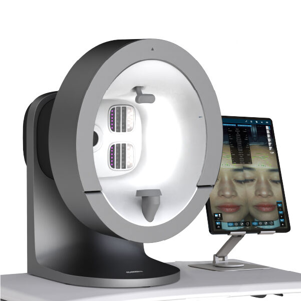
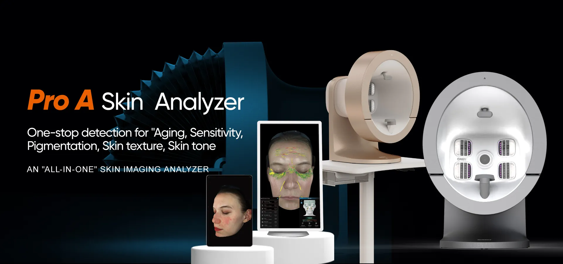
Rosacea is a chronic, progressive inflammatory disorder that manifests along a spectrum of subtypes, each with distinct clinical features and treatment needs. From the vascular flushing and redness of erythematotelangiectatic rosacea (ETR) to the papules and pustules of papulopustular rosacea (PPR), misclassifying subtypes can lead to ineffective therapies (e.g., antibiotics prescribed for vascular rosacea) or disease progression. MEICET’s Pro-A Skin Imaging Analyzer, with its multi-spectral imaging capabilities, provides clinicians with the detailed data needed to distinguish subtypes, ensuring targeted care that addresses root causes and prevents escalation.
Identifying Vascular vs. Inflammatory Features
Rosacea’s subtypes are defined by their dominant features—vascular, inflammatory, or a combination—and the Pro-A’s imaging modes isolate these characteristics with precision:
- Polarized light imaging highlights vascular dilation, the hallmark of ETR. It visualizes fine telangiectasias (dilated blood vessels) and persistent erythema (redness) that spares sun-protected areas, distinguishing ETR from other conditions. In polarized mode, ETR appears as a network of red lines (telangiectasias) overlaying diffuse redness—signaling the need for vascular lasers, brimonidine gel, or anti-inflammatory topicals (e.g., azelaic acid).
- UV imaging detects porphyrins, fluorescent byproducts of Cutibacterium acnes activity, which are linked to PPR. Elevated porphyrin levels in UV mode indicate microbial involvement, confirming PPR and guiding antibiotic therapy (topical or oral) or antimicrobial agents (e.g., metronidazole).
- RGB imaging maps papules (solid bumps) and pustules (pus-filled lesions), which are exclusive to PPR. These lesions appear as raised, well-defined structures in RGB mode, distinguishing PPR from ETR, which lacks such features.
Consider a patient presenting with facial redness and a history of “breakouts.” Pro-A scans reveal:
- Polarized light: widespread telangiectasias and diffuse redness (vascular features).
- UV imaging: moderate porphyrin fluorescence (microbial activity).
- RGB imaging: scattered papules on the cheeks (inflammatory features).
This confirms mixed rosacea (ETR + PPR), guiding a combined plan: vascular laser to address telangiectasias, topical metronidazole to reduce porphyrins, and azelaic acid to calm inflammation—ensuring all active components are treated.
Distinguishing Overlap and Atypical Presentations
Many patients have overlapping subtypes or atypical features, requiring nuanced diagnosis. The Pro-A’s integrated analysis measures each component, ensuring no aspect is overlooked:
- Phymatous rosacea, a rare subtype characterized by skin thickening (e.g., rhinophyma), presents with RGB evidence of irregular texture and thickening, paired with polarized light vascular changes. This subtype requires isotretinoin or surgical revision alongside anti-inflammatory treatments.
- Ocular rosacea, which affects the eyes, may correlate with facial findings: Pro-A scans showing ETR on the cheeks often accompany ocular symptoms (e.g., dryness, redness), prompting referral to an ophthalmologist.
- Atypical rosacea in darker skin tones may present with post-inflammatory hyperpigmentation (PIH) masking redness. Pro-A scans reveal polarized light vascular changes and UV porphyrins beneath the pigment, confirming rosacea and guiding treatments to address both inflammation and PIH.
A patient with dark skin and “facial bumps” undergoes Pro-A scanning:
- RGB shows pustules and PIH (masking redness).
- Polarized light reveals underlying vascular dilation.
- UV imaging shows high porphyrin levels.
This confirms PPR with PIH, guiding a plan of topical antibiotics (to reduce porphyrins), anti-inflammatories (to calm vascularity), and brighteners (to address PIH)—avoiding misdiagnosis as acne.
Tracking Response to Targeted Therapies
Rosacea management requires ongoing adjustment, as subtypes can evolve over time (e.g., ETR progressing to PPR). The Pro-A’s follow-up scans measure treatment success objectively:
- For ETR patients on vascular laser therapy, polarized light scans track reduced telangiectasia density and erythema—confirming whether the laser is effectively targeting vasculature.
- PPR patients using antibiotics show reduced UV porphyrin fluorescence and RGB papule count, indicating microbial activity is controlled. This guides when to transition to maintenance topicals (e.g., azelaic acid) to avoid antibiotic resistance.
- Patients with mixed subtypes have combined metrics: reduced polarized light redness (vascular response) and UV porphyrins (inflammatory response) confirming the treatment plan is addressing both components.
A patient with ETR treated with brimonidine gel has later Pro-A scans showing reduced polarized light redness—confirming the medication is working. When follow-up scans much later reveal new papules and increased UV porphyrins (PPR development), the clinician adds topical metronidazole—adapting to the evolving subtype.
The Pro-A’s multi-spectral imaging transforms rosacea diagnosis from a subjective assessment to a data-driven process. By distinguishing subtypes, measuring overlap, and tracking response, it empowers clinicians to deliver targeted, effective care that prevents progression, reduces symptoms, and improves quality of life for patients with this challenging condition.