 EN
EN
 AR
AR
 BG
BG
 HR
HR
 CS
CS
 DA
DA
 NL
NL
 FI
FI
 FR
FR
 DE
DE
 EL
EL
 HI
HI
 IT
IT
 JA
JA
 KO
KO
 NO
NO
 PL
PL
 PT
PT
 RO
RO
 RU
RU
 ES
ES
 SV
SV
 TL
TL
 IW
IW
 ID
ID
 SR
SR
 SK
SK
 SL
SL
 UK
UK
 VI
VI
 SQ
SQ
 HU
HU
 TH
TH
 TR
TR
 FA
FA
 AF
AF
 MS
MS
 UR
UR
 BN
BN
 LA
LA

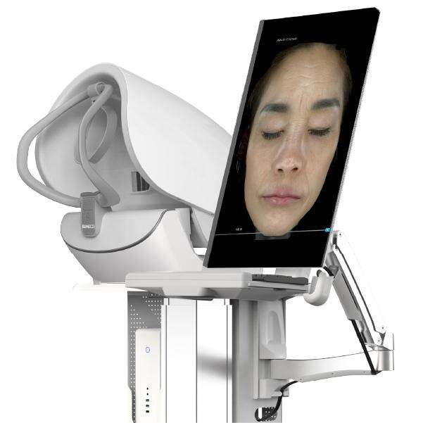
Facial rejuvenation requires precision that transcends surface-level assessments. Success depends on understanding how volume distribution, skin laxity, and underlying tissue structure interact—a complexity that basic imaging struggles to capture. MEICET’s MC88 Full Facial Skin Analyzer redefines this process, using high-definition multi-spectral imaging and tissue density analysis to map facial contours, and guide rejuvenation strategies with anatomical accuracy. For dermatologists and aesthetic practitioners, this technology transforms pre-procedural planning from educated guesswork to a data-driven science.
Mapping Volume and Contour with Anatomical Precision
The human fac今e is a dynamic landscape of curves and planes, where subtle asymmetries or hidden structural changes can impact rejuvenation outcomes. The MC88’s advanced imaging captures this complexity by creating detailed, layered visualizations that reveal:
- Regional volume characteristics in areas such as the mid-cheeks, temples, or jawline. Unlike standard photos, which flatten depth, MC88’s multi-angle imaging (frontal, lateral, oblique) quantifies how volume distribution in one area (e.g., the cheeks) influences contours in adjacent zones (e.g., the lower eyelids). This holistic view prevents over-focusing on one region at the expense of overall facial harmony.
- Skin laxity patterns by tracking how skin texture and elasticity vary across facial zones. For a patient seeking jawline refinement, MC88 scans may reveal that laxity in the lower face (not just volume changes) contributes to a “soft” appearance. This insight prompts a targeted approach: addressing laxity with collagen-stimulating treatments alongside strategies to enhance contour, rather than relying on a single modality that might yield unnatural results.
- Facial symmetry in ways basic imaging misses. A patient with a slightly fuller left cheek may appear balanced in standard photos, but MC88’s quantitative analysis measures differences in tissue density and contour that would become noticeable post-rejuvenation. By quantifying these asymmetries, clinicians can adjust treatment approaches—allocating more attention to the right side to achieve natural balance.
Consider a patient pursuing mid-face rejuvenation to address “hollowness.” Standard photos might suggest uniform volume changes, but MC88’s layered imaging reveals that the deficit is concentrated in the lateral cheeks, with the medial cheeks retaining more volume. This guides precise targeting of the lateral zone, avoiding over-treatment that would widen the face unnaturally. The result is a lifted, youthful contour that respects the patient’s natural anatomy.
Informing Planning with Quantitative Tissue Analysis
One of the MC88’s most valuable features is its ability to analyze tissue layers and density, providing objective data to guide rejuvenation strategies. This collaborative tool allows clinicians and patients to explore possibilities while setting realistic expectations:
- A patient desiring lip enhancement can review MC88’s lip tissue density maps, which illustrate how structural characteristics influence contour and harmony with the nose and chin. Based on tissue density gradients, the analysis may highlight that enhancing natural lip borders (rather than pursuing uniform fullness) better aligns with the lip’s anatomical boundaries, preventing a “disconnected” appearance.
- For mid-face rejuvenation, MC88’s subcutaneous layer analysis identifies key structural supports by mapping fat pad distribution and depth. It highlights how targeting the deep medial cheek fat pad (a natural support structure) can enhance mid-face elevation, which in turn softens lower eyelid contours and reduces nasolabial folds. Patients gain clarity on how addressing improves overall facial balance, not just surface appearance.
- Jawline refinement with MC88 focuses on bone-tissue interface mapping to identify stable anatomical landmarks. A patient with a weak mandible may learn, through density metrics, how structural characteristics influence contour; the data may suggest combining collagen-stimulating treatments with strategies to enhance the mandible’s natural axis, creating a more cohesive jawline.
This transparency is critical for patient trust. When a patient understands, via MC88’s tissue maps, that achieving “defined cheekbones” relies on enhancing the natural curve of the zygomatic arch (rather than creating an artificial peak), they are more likely to embrace a plan that enhances their unique features. It also reduces post-procedural dissatisfaction, as patients gain clear awareness of anatomical limitations and possibilities.
Supporting Post-Procedural Evaluation
Imaging doesn’t end with the procedure. The MC88’s follow-up scans quantify how tissue responds to rejuvenation over time, ensuring both safety and longevity:
- Contour consistency checks for uneven settling, which can create textural irregularities. A patient with mid-face rejuvenation might have MC88 scans at one month that reveal slight textural differences in the cheek area. This early detection allows for targeted skincare adjustments to support uniform healing, avoiding the need for more invasive interventions later.
- Tissue response is assessed via multi-spectral texture analysis, which detects inflammation or changes in elasticity. Raised areas may signal excessive collagen formation, while indentations could indicate uneven tissue response—requiring targeted soothing or adjustment of subsequent treatments.
For clinicians, this means moving beyond “how it looks” to “how it functions”—ensuring rejuvenation treatments are both aesthetic and anatomically sound. The MC88 transforms facial rejuvenation from static interventions to dynamic processes, where ongoing multi-spectral monitoring ensures results evolve naturally with the patient’s anatomy.