 EN
EN
 AR
AR
 BG
BG
 HR
HR
 CS
CS
 DA
DA
 NL
NL
 FI
FI
 FR
FR
 DE
DE
 EL
EL
 HI
HI
 IT
IT
 JA
JA
 KO
KO
 NO
NO
 PL
PL
 PT
PT
 RO
RO
 RU
RU
 ES
ES
 SV
SV
 TL
TL
 IW
IW
 ID
ID
 SR
SR
 SK
SK
 SL
SL
 UK
UK
 VI
VI
 SQ
SQ
 HU
HU
 TH
TH
 TR
TR
 FA
FA
 AF
AF
 MS
MS
 UR
UR
 BN
BN
 LA
LA

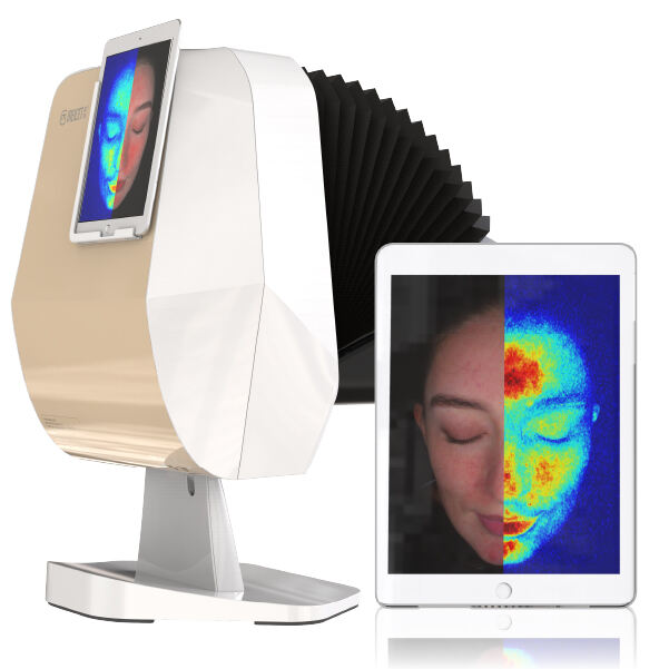
Facial fillers have become indispensable in aesthetic medicine, offering versatile solutions for volume restoration, contour enhancement, and wrinkle reduction. However, their success hinges on more than precise injection technique: clinicians must ensure the filler integrates smoothly with surrounding tissue, avoids migration, and maintains natural contours over time. Traditional assessments, reliant on visual inspection and palpation, often miss subtle signs of uneven settling, inflammation, or early migration—risks that can compromise results or trigger adverse events. MEICET’s MC88 Skin Analyzer addresses this gap by using advanced imaging to evaluate post-filler tissue response, providing clinicians with the detailed insights needed to ensure both safety and aesthetic success.
Assessing Filler Distribution and Contour Harmony
Even filler distribution is critical for natural results, as clumping or migration can lead to lumps, asymmetry, or an artificial appearance. The MC88’s high-resolution imaging identifies these issues early, allowing for timely intervention:
- Polarized light imaging highlights the interface between filler and surrounding tissue, revealing whether the product is evenly dispersed or concentrated in pockets. For example, a patient soon after cheek filler may have polarized light scans showing uniform highlights across the treated area—confirming proper placement. In contrast, a jawline scan with clustered, irregular highlights signals localized accumulation, prompting gentle massage to redistribute the filler before it solidifies.
- RGB imaging maps surface contours, comparing treated areas to adjacent regions to ensure seamless transitions. A patient with lip fillers should have smooth, gradual blending between the lips and surrounding skin; RGB scans showing abrupt elevation or unevenness may indicate over-filling, guiding targeted dissolution of excess product to restore natural proportions.
- UV imaging indirectly assesses tissue health around the filler by detecting changes in barrier function. Increased UV fluorescence in treated areas can signal subclinical inflammation, which may disrupt integration. This finding guides adjustments to aftercare, such as adding soothing serums or avoiding irritants (e.g., retinoids) until the skin stabilizes.
Consider a patient a short time after chin filler: MC88 scans reveal polarized light highlights indicating slight migration toward the neck, RGB showing a subtle step-off between the chin and jawline, and UV with mild fluorescence (suggesting early inflammation). The clinician can intervene with targeted massage to reposition the filler, recommend a calming moisturizer to address inflammation, and schedule a follow-up scan soon after to confirm resolution—preventing visible irregularities and ensuring the filler maintains its intended contour.
Detecting Early Signs of Complications
While serious complications from fillers are rare, early detection of issues like granulomas, vascular compromise, or infection is critical to minimizing damage. The MC88’s detailed imaging flags subtle warning signs that might escape visual inspection:
- Texture irregularities in RGB mode, such as small, firm raised areas, may indicate incipient granulomas—immune reactions to the filler. These appear as distinct, well-circumscribed bumps that differ from normal post-injection swelling, which is diffuse and soft. Early detection allows for prompt intervention with steroid injections, preventing granuloma progression and preserving results.
- Vascular changes in polarized light imaging, such as localized blanching (pale areas) or increased redness, can signal reduced blood flow—a precursor to tissue ischemia. This is particularly critical in high-risk areas like the nose or tear troughs, where vascular compromise can lead to scarring. MC88 scans soon after injection provide a baseline, enabling clinicians to compare subsequent scans and act quickly if vascular changes emerge.
- Asymmetric volume in comparative scans (pre- vs. post-treatment) may reveal uneven absorption or migration, guiding touch-ups to maintain balance. For example, a patient with cheek fillers may have scans showing more volume on one side, prompting a small touch-up to the other to restore symmetry.
In one clinical scenario, a patient a few days after tear trough filler presents with mild swelling. MC88 scans show polarized light highlighting a localized pale area (suggesting vascular compression) and RGB confirming a firm nodule. The clinician recognizes these as early signs of vascular compromise, administers hyaluronidase to dissolve the problematic filler, and monitors with follow-up scans—preventing tissue necrosis and scarring.
Guiding Long-Term Maintenance Plans
Fillers degrade over time due to enzymatic activity, but their rate of absorption varies by patient, product, and injection site. The MC88 helps clinicians determine optimal touch-up timing by tracking volume and tissue response:
- Consistent scans over time track how much filler remains versus natural tissue. A patient with significant volume may benefit from a touch-up to maintain results, while one with more gradual absorption might need a full treatment to restore desired volume.
- Tissue response in polarized light imaging indicates whether the filler is integrating with surrounding tissue or being encapsulated. Encapsulation appears as a distinct “halo” around the filler in polarized mode, signaling the need to switch to a different product (e.g., a more cohesive hyaluronic acid) for future treatments to reduce encapsulation risk.
- Patient-specific factors—such as metabolism, skin thickness, and activity level—are correlated with scan data to personalize maintenance intervals. Patients with faster metabolism (evident in quicker filler degradation on scans) may need more frequent touch-ups, while those with thicker skin (slower degradation) can extend intervals.
For a patient with jawline fillers, MC88 scans over time show steady volume with no signs of encapsulation or inflammation. This confirms the product is integrating well, justifying an extended interval for the next touch-up—balancing efficacy, patient convenience, and cost-effectiveness.
The MC88 Skin Analyzer elevates post-filler care from reactive to proactive, ensuring clinicians have the data to deliver safe, natural, and long-lasting results. By evaluating distribution, detecting complications early, and guiding maintenance, it transforms filler procedures into refined, patient-specific interventions that enhance facial harmony without compromising safety.